
Advanced Eye Care Technology in Huntington Beach
Most of what threatens your vision starts quietly. We invest in imaging that finds those changes years before you would notice them, and that lets us compare your eyes to themselves over time rather than guessing.
Optos California
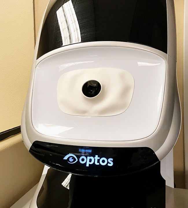
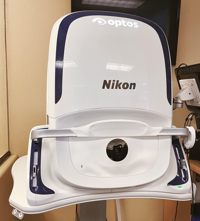
A 200−degree view of your retina in a single image, usually without dilation drops.
How It Works
A scanning laser captures over 80% of your retina in one shot. A standard view shows roughly a quarter of that, a section at a time. Each image is stored, so at your next visit we compare pictures rather than memories.
Why It Matters
Retinal tears, detachments, and tumors tend to start in the far periphery — the part a standard view is most likely to miss. The retina is also the only place we can see blood vessels directly, so the image sometimes shows early signs of high blood pressure or diabetes before other symptoms appear. And in most cases, you can skip the dilation drops and drive yourself home.
Zeiss Optical Coherence Tomography (Oct)
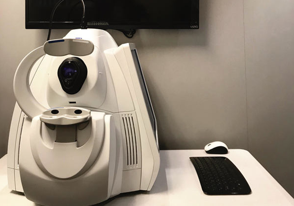
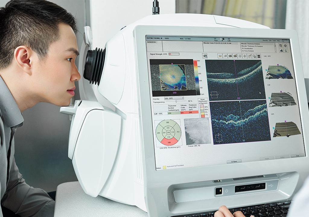
A detailed scan of the layers beneath the surface of your retina
How It Works
Like an ultrasound, but using light instead of sound. It measures reflections to build a cross-section of your retina, layer by layer, down to a few microns.
Why It Matters
Reveals glaucoma damage, macular degeneration, and diabetic swelling years before you'd notice a change in your vision. Takes seconds, no drops or dye required.
Humphrey Visual Field Analyzer
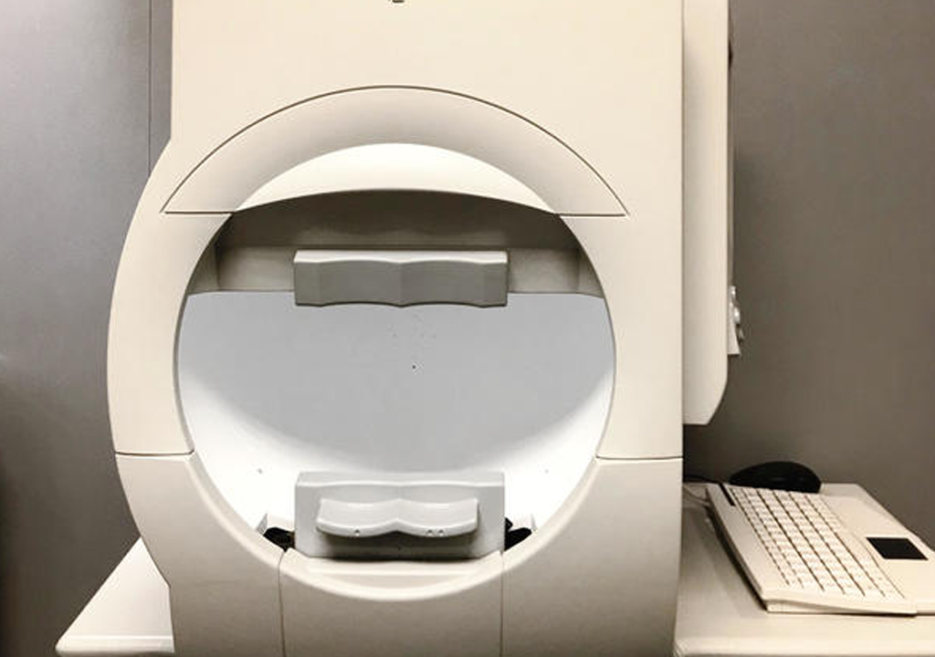
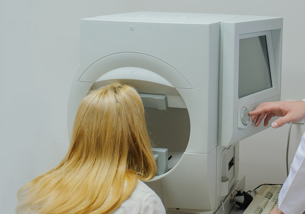
Measures how well you see at every point across your field of vision.
How It Works
You look straight ahead and press a button each time you notice a faint light flicker at the edge of your vision. Point by point, that builds a map of where your vision is strong and where it's thinning.
Why It Matters
Glaucoma takes peripheral vision so gradually that the brain fills in the gap, and people often lose a great deal before noticing. This test finds that loss while it is still invisible to you.
Not just for glaucoma
Visual field testing shows us how the entire visual pathway is functioning, so we use it well beyond glaucoma care.
Neurological conditions. The nerve pathway from your eyes to the back of your brain is long, and damage anywhere along it shows up as a specific pattern of missing vision. Because the pattern points to the location, a visual field can occasionally be the first clue that something needs imaging — and after a stroke, aneurysm, or brain surgery, it documents exactly what vision remains, which matters for driving, reading, and rehabilitation.
Optic nerve conditions. The optic nerve can be damaged by reduced blood flow (AION), by inflammation (optic neuritis, sometimes the first sign of multiple sclerosis), or by pressure — either from swelling inside the skull or from deposits called drusen within the nerve itself. The visual field shows how much function has been lost and whether it's stable, improving, or progressing. For conditions we co-manage with a neurologist or neuro-ophthalmologist, these results go directly to them.
Eyelid surgery evaluations. When drooping upper eyelids block your upper field of vision, insurance generally requires documentation before approving surgery. If requested by your surgeon, we perform the visual field testing that insurers need. Medication monitoring. Hydroxychloroquine (Plaquenil) and a few other long-term medications carry a small risk of retinal toxicity. Guidelines call for a baseline exam and annual screening, which we perform alongside OCT imaging. If you take one of these medications, we coordinate directly with your prescribing physician.
Referred by another doctor? Send us the request and we'll get the results back to them promptly.
MYAH
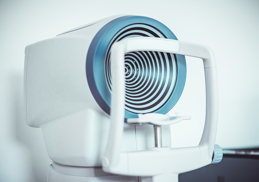
One instrument, three measurements: the shape of your cornea, the length of your eye, and the health of your oil glands.
Corneal Topography
How It Works
Maps the exact shape of the front surface of your eye, point by point.
Why It Matters
Detects keratoconus early, and maps the surface we fit specialty contact lenses to.
Axial Length Interferometry
How It Works
Measures the front-to-back length of the eye to a fraction of a millimeter.
Why It Matters
Meibography
An infrared photograph of the inside of your eyelids, which makes the oil glands visible in a single image.
Why It Matters
Shows whether your glands are healthy, blocked or already lost, which is what determines the treatment. We turn the screen around and show you.
USING THEM TOGETHER
No single instrument tells the whole story. The value is in combining them.
Fitting specialty contact lenses
The MYAH maps the shape of your cornea; the anterior segment OCT measures the space between a lens and your eye. Together they tell us how a scleral lens will actually sit before we have put one on your eye, which means fewer trial lenses, fewer visits, and a better fit for the patients who need sclerals precisely because their corneas are not a standard shape.
Watching for glaucoma
Structure and function do not always decline together. The OCT shows us the physical state of your optic nerve; the visual field shows us how well it is working. Damage can show up in either one first, so we follow both over time rather than relying on a single number.
Managing dry eye
Meibography shows us the condition of your oil glands. Tear film measurements show us what is actually reaching the surface of your eye. The combination tells us whether your dry eye is a production problem or an evaporation problem, which is what determines the treatment.

© 2026 Beachside Optometry. All rights Reserved -
Accessibility Statement
-
Privacy Policy
-
Sitemap
Managed and Designed by 